The eyes are among the most delicate and rapidly deteriorating organs in the body. A condition that causes mild irritation in the morning can progress to a corneal ulcer or vision-threatening infection by evening if left unaddressed. Yet eye problems in pets are also among the most commonly dismissed — redness and discharge are frequently attributed to dust or allergies and treated with home remedies, when they may be signs of something that requires immediate veterinary intervention.
In India, several environmental factors compound the risk significantly. Year-round dust and air pollution irritate the ocular surface. High humidity during monsoon promotes the growth of bacterial and fungal pathogens. Dense urban populations of stray animals increase the transmission of contagious eye infections. And a large population of brachycephalic (flat-faced) breeds — Pugs, Persian cats, Bulldogs, Shih Tzus — carry anatomical vulnerabilities that make them prone to chronic eye disease from the day they are born.
Breed-Related Eye Vulnerability
Brachycephalic breeds — inherently higher risk
Pugs, Bulldogs, Shih Tzus, Pekingese, Boston Terriers, Persian cats, and Himalayan cats have shallow eye sockets that cause the eye to protrude further than in other breeds. This reduces the eyelid's ability to fully cover and protect the cornea, leads to reduced blink reflex, and causes the corneal surface to dry out faster. These breeds are significantly more prone to corneal ulcers, dry eye, pigmentary keratitis, and exposure keratopathy. They require more frequent eye cleaning and proactive veterinary eye checks — at least annually, ideally every 6 months.
Large-breed dogs — Labrador Retrievers, German Shepherds, Golden Retrievers — are more prone to entropion (inward-rolling eyelids) and hereditary cataracts. Senior dogs of any breed are at increased risk of progressive retinal atrophy, nuclear sclerosis, and age-related cataracts. Understanding your pet's breed-specific vulnerabilities allows you to monitor for early signs and catch problems before they become irreversible.
Common Eye Conditions in Indian Dogs & Cats
👁️ Conjunctivitis (Pink Eye)
Inflammation of the conjunctiva — the thin membrane lining the eyelids and the white of the eye. The most frequently diagnosed eye condition in Indian pets. Causes include bacterial infections (especially Staphylococcus and Streptococcus), viral infections (distemper, feline herpesvirus), allergies, dust, and smoke. Signs include redness, swelling, excessive blinking, and watery or coloured discharge. Viral conjunctivitis is contagious between cats and requires isolation of affected animals.
🔴 Corneal Ulcers
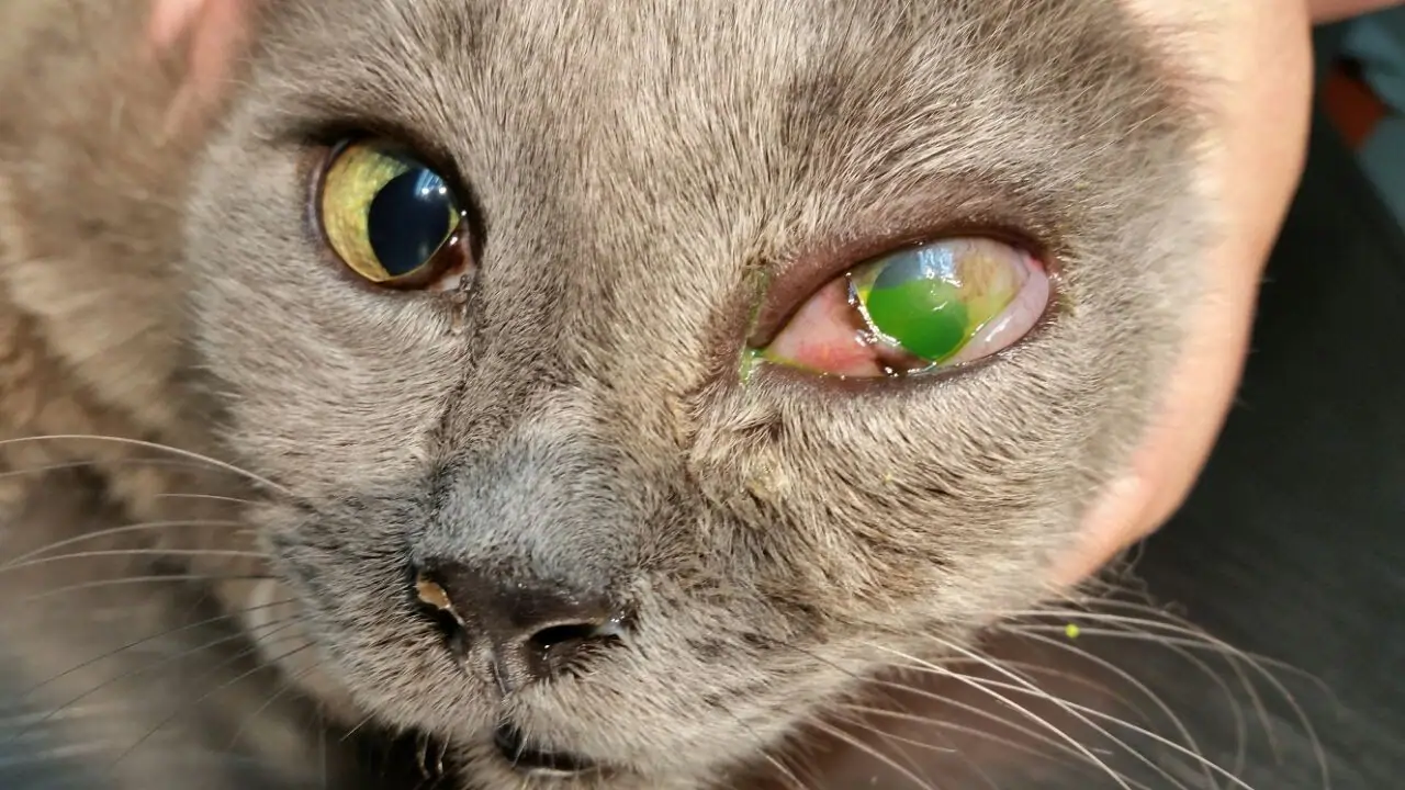
Open wounds on the corneal surface — extremely painful and capable of progressing to corneal perforation within 24–48 hours if untreated. Causes include trauma (scratch from another animal, foreign body), untreated dry eye, chemical splash, or deep infections. The dog or cat will squint, keep the eye partially or fully closed, paw at the face, and avoid light. Never apply corticosteroid eye drops to an eye with a corneal ulcer — steroids impair healing and can convert a superficial ulcer to a deep, perforating one. Fluorescein stain at the vet clinic confirms the ulcer.
💧 Dry Eye (KCS)
Keratoconjunctivitis sicca occurs when the lacrimal glands fail to produce sufficient tears, leaving the cornea chronically under-lubricated. Most commonly immune-mediated in dogs; also caused by certain antibiotics (sulphonamides) and systemic diseases. Signs include thick, yellow-green mucoid discharge, dull corneal surface, and recurrent conjunctivitis. Without treatment, the cornea becomes pigmented and scarred, leading to permanent vision impairment. KCS is managed lifelong with tear stimulant eyedrops (cyclosporine or tacrolimus) and artificial tear supplementation.
☁️ Cataracts
Opacity of the crystalline lens, causing a blue-white cloudiness and progressive vision loss. Causes include genetics (hereditary cataracts in Labrador Retrievers, Poodles, Cocker Spaniels), diabetes mellitus (diabetic cataracts progress very rapidly), ageing, and previous ocular inflammation. Note: nuclear sclerosis — a bluish haziness of the lens visible in dogs over 7 years — is often mistaken for cataracts but does not significantly impair vision and does not require surgical treatment. A veterinary ophthalmology examination distinguishes the two. Surgical correction (phacoemulsification) is available in India at specialist centres.
⚡ Glaucoma
Pathologically elevated intraocular pressure (IOP) that causes severe pain, rapid optic nerve degeneration, and irreversible vision loss. Primary glaucoma is heritable in certain breeds (Cocker Spaniels, Basset Hounds, Beagles). Secondary glaucoma follows uveitis (internal eye inflammation), lens luxation, or intraocular tumours. Acute signs include sudden redness, a visibly enlarged eye, corneal cloudiness, severe pain (pawing, reluctance to be touched around the head), and vision loss. Tonometry (IOP measurement) at the clinic confirms the diagnosis. Treatment involves reducing IOP with medication and, in some cases, surgery to preserve the remaining eye.
🍒 Cherry Eye
Prolapse of the third eyelid gland — a red, rounded, fleshy mass visible at the inner corner of the eye, resembling a small cherry. Common in young dogs, particularly Bulldogs, Beagles, Cocker Spaniels, and mixed breeds. Cherry eye is not just cosmetic: the prolapsed gland produces approximately 30–40% of the eye's total tear film. Left untreated, it leads to chronic dry eye. Surgical repositioning (not removal) of the gland is the recommended treatment — removal eliminates that portion of tear production permanently.
The Third Eyelid — What It Tells You
Dogs and cats have a nictitating membrane (third eyelid) — a pale, pinkish-white tissue at the inner corner of each eye. Under normal circumstances it is barely visible. When it becomes prominently visible — covering part of the eye surface — it is almost always a sign that something is wrong, either locally with the eye or systemically in the rest of the body.
- Local causes: Eye pain or irritation, Horner's syndrome (neurological), conjunctivitis, foreign body under the eyelid
- Systemic causes: Dehydration, severe weight loss, systemic illness (particularly in cats with "Haw's syndrome" — bilateral third eyelid protrusion seen with gastrointestinal disease), tetanus
Warning Signs — Know When to Act
The following symptoms should always prompt veterinary evaluation. The ones marked urgent require same-day attention:
Daily Eye Care for Pet Parents
Routine eye care is simple and takes less than two minutes — but done consistently, it allows you to detect early changes before they become serious. It also keeps the eye surface clean of discharge that can harbour bacteria and irritate the delicate periocular skin.
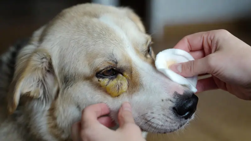
- Gentle daily cleaning — Use a clean, damp cotton ball or a veterinarian-recommended sterile eye wipe to gently remove discharge from the inner corner of each eye. Always wipe outward — never inward across the eye surface. Use a separate cotton ball for each eye.
- Trim hair around the eyes — Particularly important in Shih Tzus, Lhasa Apsos, Schnauzers, and long-haired cats. Hair touching the cornea acts as a continuous foreign body irritant, causing chronic conjunctivitis and corneal scratching. Use blunt-nosed scissors or ask a groomer.
- Avoid environmental irritants — Keep your pet away from smoke, chemical fumes, construction dust, and strong cleaning products. When travelling by car, keep windows only partially open — wind-driven particles commonly cause corneal irritation and foreign bodies.
- Maintain parasite control — Ticks can occasionally attach near the eye; fleas cause irritation that leads to eye-rubbing. Year-round prevention reduces these risks.
- Check discharge daily — A small amount of clear or slightly white discharge at the inner corner after sleep is normal. Any change in colour, consistency, quantity, or associated redness is a reason to contact your vet.
- Annual eye checks for all pets; every 6 months for at-risk breeds — Many early eye conditions — early KCS, early cataracts, early entropion — are identified at routine health checks before they cause symptoms. Do not wait for your pet to show obvious signs before having their eyes examined.
Frequently Asked Questions
Is eye discharge normal in dogs and cats?
A small amount of clear or slightly white discharge collected at the inner corner of the eye after sleep is generally normal — this is the eye's way of clearing debris from the night. However, thick, yellow, green, grey, or bloody discharge at any time of day, or any discharge accompanied by redness, squinting, or swelling, is not normal and requires veterinary evaluation.
Can I use human eye drops for my pet?
No. Human eye drops — including OTC artificial tears, antihistamine drops, and antibiotic drops — are formulated for human eye pH, tonicity, and physiology. Many contain preservatives or drug concentrations that can damage the canine or feline cornea. Plain preservative-free sterile saline can be used to flush debris from the eye, but any medicated product requires a veterinary prescription and specific diagnosis.
My dog's eyes look blue or cloudy — is this cataracts?
Not necessarily. Nuclear sclerosis — a normal age-related change in the lens that produces a blue-grey haze — is extremely common in dogs over 7 years old and does not significantly impair vision. It does not require treatment. Cataracts produce a denser, whiter opacity and do affect vision. A veterinary examination, including menace response and pupillary light reflex testing, distinguishes the two — a definitive answer requires an ophthalmology-grade fundic examination.
Can eye infections spread between pets?
Yes — certain causes of conjunctivitis are contagious between animals of the same species. Feline herpesvirus (FHV-1) is highly contagious between cats and is the most common cause of conjunctivitis in multi-cat households. Canine distemper virus causes conjunctivitis in dogs. If one pet in a multi-pet household develops conjunctivitis, isolate the affected animal and have all pets evaluated by a veterinarian before assuming the cause is non-contagious.
Conclusion
Eye problems in dogs and cats share a common characteristic: they rarely improve on their own and frequently worsen quickly. The anatomy of the eye means that inflammation, infection, and pressure elevation cause damage in hours, not days — and that damage is often irreversible. The conditions covered in this guide range from manageable chronic diseases to acute emergencies, but all share the same lesson: early recognition and prompt veterinary care produce dramatically better outcomes than delayed treatment.
Build a simple daily eye check into your routine — take thirty seconds to look at each eye for discharge, redness, or asymmetry. Know which signs require a same-day call to your veterinarian. And if your pet is a flat-faced breed, treat eye health as a proactive, scheduled priority rather than something to address only when a problem becomes visible. Your pet cannot tell you their eye hurts — but the signs are always there if you know what to look for.
Related Guides
This content is provided for educational purposes only and is not a substitute for professional veterinary advice, diagnosis, or treatment. Eye conditions in pets can deteriorate rapidly — if your pet shows signs of eye pain, sudden vision change, or acute redness, seek veterinary care the same day. Never apply any eye medication without a veterinary prescription and confirmed diagnosis.