Joint disease is one of the most underdiagnosed conditions in Indian pets. Unlike a wound or a bout of vomiting, joint pain develops gradually and is easy to miss — partly because dogs and cats instinctively conceal pain and discomfort, and partly because owners often attribute reduced activity or stiffness to "getting old" rather than to a treatable medical condition. By the time most pets are diagnosed with osteoarthritis in India, the disease has been present and progressive for months to years.
This matters because joint disease is not simply a quality-of-life issue — it is a pain management issue. A dog that struggles to climb stairs, a cat that stops jumping to favourite spots, a large breed that hesitates before lying down: these are animals in daily pain. The good news is that with early recognition, appropriate management, and veterinary partnership, most pets with joint disease can maintain active, comfortable lives well into their senior years.
Common Joint Conditions in Dogs & Cats
Osteoarthritis (OA)
The progressive breakdown of articular cartilage — the smooth tissue that cushions bones at their contact points. As cartilage degrades, bone surfaces rub together, causing inflammation, pain, and the formation of osteophytes (bone spurs). OA affects approximately 20% of dogs over 1 year old and is nearly universal in dogs over 11. In cats it is severely underdiagnosed — studies suggest over 90% of cats over 12 have radiographic evidence of OA, yet most owners are unaware. OA is not curable, but it is highly manageable.
Hip Dysplasia
Abnormal development of the hip joint where the femoral head does not seat properly in the acetabulum, causing joint laxity, cartilage damage, and eventual severe OA. Strongly heritable — responsible breeding and OFA/PennHIP screening can reduce incidence. Seen frequently in Labrador Retrievers, German Shepherds, Golden Retrievers, and large Indian breeds including Rajapalayam and Mudhol Hounds. Rapid growth, excessive dietary calcium, and overexercise in puppies worsen genetically susceptible individuals.
Elbow Dysplasia
An umbrella term for several developmental conditions affecting the elbow joint: fragmented coronoid process, osteochondrosis dissecans (OCD), and ununited anconeal process. All cause incongruity of the elbow joint surfaces, leading to chronic pain, lameness — typically presenting as a front-limb limp in young large-breed dogs — and secondary OA. Diagnosis requires radiographs and often CT scanning. Surgical treatment gives significantly better outcomes when undertaken early.
Cranial Cruciate Ligament (CCL) Rupture
The canine equivalent of an ACL tear — the most common orthopaedic injury in dogs. Unlike sports injuries in humans, CCL rupture in dogs is usually the end-stage of chronic, progressive ligament degeneration rather than a single traumatic event. Affected dogs show sudden hind-limb lameness and the stifle (knee) shows a characteristic "drawer sign" on examination. Without surgical stabilisation, the joint becomes severely arthritic. Three proven surgical techniques exist: TPLO, TTA, and lateral suture — your vet will advise based on breed, size, and activity.
Patellar Luxation
Medial patellar luxation — where the kneecap slips out of its groove towards the inside of the leg — is the most common orthopaedic condition in small-breed dogs in India. Spitz, Pomeranian, Poodle, and Shih Tzu breeds are particularly predisposed. Affected dogs show intermittent "skipping" lameness where they lift the affected leg for a few steps. Severity is graded 1–4; grades 3–4 require surgical correction to prevent permanent joint damage.
Spondylosis & IVDD
Spondylosis deformans — bony bridges forming between vertebrae — is extremely common in senior cats and often found incidentally on X-rays. It causes stiffness and reduced spinal flexibility. Intervertebral disc disease (IVDD) involves disc material herniating into the spinal canal, causing pain, weakness, or paralysis. IVDD is particularly common in chondrodystrophic dog breeds: Dachshund, Beagle, Shih Tzu, and French Bulldog. Acute disc herniation with hind-limb weakness or paralysis is a spinal emergency requiring urgent veterinary assessment.
🐕 High-Risk Breeds — Be Proactive from Puppyhood
- Large breeds: Labrador Retriever, Golden Retriever, German Shepherd, Rottweiler, Great Dane — hip/elbow dysplasia, CCL rupture, OA
- Small breeds: Spitz, Pomeranian, Chihuahua, Poodle, Shih Tzu — patellar luxation, IVDD
- Chondrodystrophic breeds: Dachshund, Beagle, French Bulldog, Basset Hound — IVDD, joint malformation
- Indian breeds: Rajapalayam, Mudhol Hound — hip dysplasia risk; Combai — joint laxity in large individuals
- Cats: Maine Coon — hip dysplasia; all cats over 10 years — high OA prevalence regardless of breed
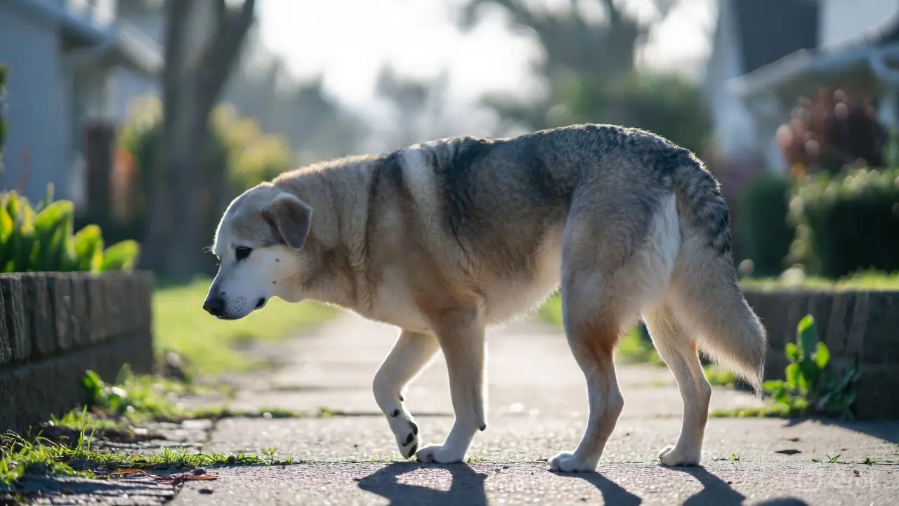
Recognising Joint Pain — Signs to Watch For
Because pets cannot tell us they are in pain, we must read their behaviour. Joint pain is consistently underreported to veterinarians because owners do not recognise the signs — particularly in cats, who show pain through withdrawal and behaviour change rather than obvious limping.
Prevention & Management Strategies
Weight Management — The Single Most Important Factor
Every extra kilogram of body weight places approximately 3–4 kg of additional force on weight-bearing joints with every step. Obesity is the single most modifiable risk factor for OA onset, progression, and pain severity. Studies show that even a 6–8% reduction in body weight produces clinically significant pain reduction in arthritic dogs. Body Condition Score (BCS) 4–5 out of 9 is the target — ribs easily felt but not visible.
Controlled, Low-Impact Exercise
The instinct to rest an arthritic pet is counterproductive — inactivity accelerates muscle loss and joint stiffness. The goal is controlled exercise that maintains muscle mass and joint lubrication without impact stress. Short, frequent walks on soft surfaces (grass, soil) are preferable to long walks on hard tiles or concrete. Swimming is ideal. Avoid ball-chasing, jumping, and sudden direction changes. "Little and often" is the guiding principle.
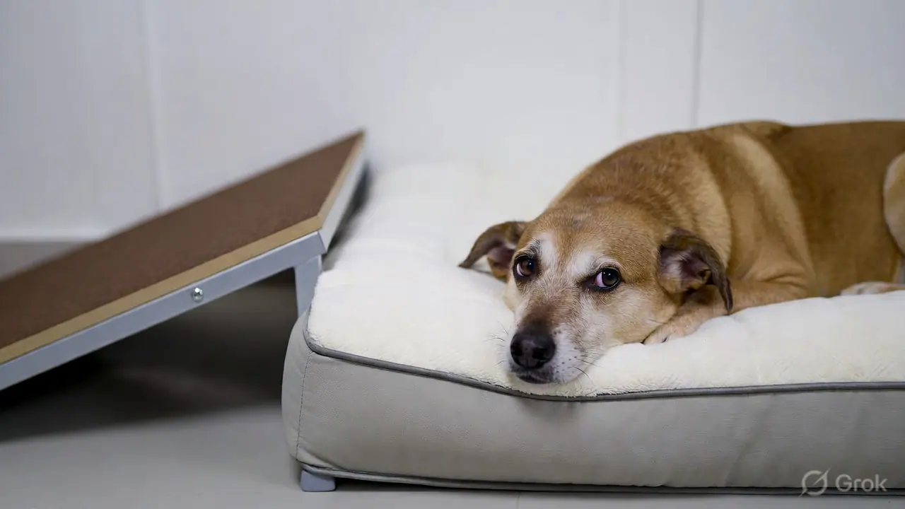
Environmental Modifications
Many Indian homes have marble or tile flooring — smooth, hard, and cold, all of which worsen joint pain and increase fall risk for arthritic pets. Place non-slip bath mats or yoga mats along movement pathways and near food bowls. Provide ramps for sofas, beds, and car access — jumping down is far more damaging than jumping up. Orthopaedic memory foam beds reduce pressure point pain significantly compared to thin cushions or bare floor.
Nutraceuticals & Joint Supplements
Several supplements have published clinical evidence for joint health — see the supplement table below. They work best started early (before or at the first signs of OA) and maintained consistently. They are not pain medications and do not replace NSAIDs in a painful, diagnosed arthritic pet — but they slow cartilage degradation and reduce inflammation, reducing the dose of medication needed over time.
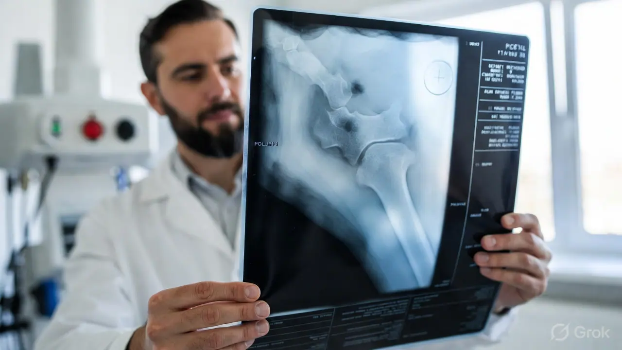
Regular Veterinary Assessments
For high-risk breeds: annual orthopaedic screening from 1 year old, including gait assessment and hip/elbow palpation. For senior pets (dogs over 7, cats over 10): biannual check-ups with mobility assessment. Radiographs confirm diagnosis and track progression. Early diagnosis opens the window for surgical intervention (dysplasia, CCL, patellar luxation) before irreversible OA develops.
Physiotherapy & Hydrotherapy
Veterinary physiotherapy — including passive range-of-motion exercises, massage, and targeted strengthening — has strong evidence for improving mobility in arthritic pets. Hydrotherapy (underwater treadmill or swimming pool) allows exercise at reduced joint loading, builds muscle without impact, and is increasingly available in larger Indian cities. Ask your vet for a referral to a certified veterinary rehabilitation practitioner.
Joint Supplements — Evidence Summary
The supplement market for pet joint health is large and inconsistent in quality. The table below summarises the compounds with the best-published evidence and gives practical guidance. Always use veterinarian-recommended formulations — quality control varies significantly between brands.
| Supplement | Mechanism | Evidence Level | Notes |
|---|---|---|---|
| Glucosamine HCl | Cartilage synthesis substrate; may reduce degradation enzymes | Moderate — most effective combined with chondroitin | Takes 4–8 weeks for full effect; safe long-term |
| Chondroitin Sulphate | Inhibits cartilage-degrading enzymes; retains water in cartilage | Moderate — synergistic with glucosamine | Often combined in one product; dose by weight |
| Omega-3 (EPA/DHA) — Fish Oil | Anti-inflammatory via prostaglandin pathway; reduces synovial inflammation | Good — among the highest evidence for OA | Use fish oil, not flaxseed (dogs convert ALA poorly); dose 75–100 mg EPA+DHA/kg body weight/day |
| Green-Lipped Mussel (Perna canaliculus) | Contains unique omega-3s, GAGs, and ETA not found in fish oil | Good — several RCTs supporting OA pain reduction | Freeze-dried powder form retains more bioactive compounds than oil extract |
| Curcumin / Turmeric extract | NF-κB pathway inhibitor; anti-inflammatory and antioxidant | Moderate — bioavailability is the key challenge | Use standardised, bioavailability-enhanced veterinary formulations — plain turmeric powder has very poor absorption |
| Boswellia Serrata | 5-LOX pathway inhibitor; reduces leukotriene-driven joint inflammation | Moderate — good safety profile; combines well with omega-3 | Widely studied in India; several veterinary products available |
| MSM (Methylsulphonylmethane) | Sulphur donor for collagen and cartilage synthesis; mild anti-inflammatory | Preliminary — often added to glucosamine/chondroitin blends | Very safe; adds sulphur missing from processed pet diets |
India-Specific Seasonal Considerations
☀️ Summer (March–June)
High ambient temperatures increase systemic inflammation, which can worsen joint pain acutely. Exercise should be limited to early morning and late evening when surfaces are cooler. Ensure excellent hydration — dehydration reduces synovial fluid volume. Tile and stone floors become dangerously slippery when the pet's paws sweat; place mats at key locations.
🌧️ Monsoon (June–September)
Humidity and barometric pressure changes are associated with increased arthritis pain in both humans and animals — the mechanism involves joint fluid pressure changes and increased nerve sensitivity. Many owners notice their arthritic pets are more painful or reluctant to move during monsoon. Maintain indoor exercise (slow leash walks inside corridors, gentle play) and keep the pet warm and dry.
❄️ Winter (November–February)
Cold is the classic arthritis trigger — muscles tighten, synovial fluid thickens, and joints become stiffer and more painful with lower ambient temperature. In North India, this effect is pronounced. Keep arthritic pets indoors and warm; use a padded, draught-free bed; warm up slowly before exercise; consider adding a lightweight vest for thin-coated dogs. Do not skip walks entirely — movement is therapeutic.
Veterinary Treatment Options
Arthritis management in veterinary medicine has advanced significantly in recent years. The multimodal approach — combining more than one type of intervention — consistently outperforms single-agent treatment. Your vet may recommend:
- NSAIDs (non-steroidal anti-inflammatory drugs) — meloxicam, carprofen, grapiprant; the cornerstone of OA pain management in dogs; require regular bloodwork monitoring for liver and kidney health; cats require feline-specific NSAIDs at much lower doses
- Gabapentin — for neuropathic (nerve-related) joint pain, particularly useful in cats and as an add-on in dogs with incomplete NSAID response
- Monoclonal antibody therapy (Librela / Solensia) — the newest advance in veterinary OA management; monthly injectable biologic that targets nerve growth factor; provides significant pain relief with a very clean safety profile; increasingly available in India
- Intra-articular injections — hyaluronic acid or platelet-rich plasma (PRP) injected directly into the joint; most useful in single-joint OA with good anatomical access
- Surgery — for CCL rupture (TPLO/TTA), hip dysplasia (juvenile pubic symphysiodesis, femoral head ostectomy, total hip replacement), patellar luxation, and elbow dysplasia; best outcomes when performed early before secondary OA becomes severe
- Acupuncture — increasingly supported by evidence for chronic musculoskeletal pain; available in several Indian metro cities through certified veterinary acupuncturists
Related Guides
This content is provided for educational purposes only and is not a substitute for professional veterinary advice, diagnosis, or treatment. Joint conditions are progressive — delayed diagnosis and treatment leads to worse long-term outcomes. If your pet shows any signs of reduced mobility, stiffness, or pain, consult your registered veterinarian promptly. Do not administer human NSAIDs (ibuprofen, aspirin, naproxen) to pets — these are toxic and can be fatal.